Scientific Program

Day 1 :

Keynote Forum

Karen Pierce

University of California, USA

Keynote: Autism from the beginning: Early screening and detection and the search for biomarkers

Time : 09:40-10:20

Biography:

Karen Pierce has been studying autism for the past 20 years and is a Leading Expert on the neural and clinical phenotype of ASD. Her research spans a range of topics from early screening and detection to eye tracking and functional magnetic resonance imaging (fMRI). Her early detection approach, called the 1-Year Well-Baby Check-Up Approach, has identified several hundred ASD toddlers around the 1st birthday and has resulted in rapid treatment access. This is a major advance because the mean age of detection in the US is around 4 years in age. She has been invited as a Keynote Speaker on the topic of Autism at both national and international conferences. Her work is published in high-impact journals and has been highlighted in the public media including CNN, The Wall Street Journal, and Time Magazine. Her research is funded by both NIH as well as private organizations such as the National Foundation for Autism Research. She has been honored by several awards including US Department of Health and Human Services IACC Top Research Paper Award, Autism Speaks Top 10 Research Paper Award, and the San Diego Health Hero Award

Abstract:

Our understanding of autism spectrum disorder (ASD) has advanced tremendously across the past decade, with a growing appreciation of its probable prenatal origins and wide-ranging biological features. Still it remains behaviorally identified and diagnosed with the mean of detection in America after around age of 3-4 years. The first three years of life are the most primary transformative period for human postnatal brain development. Such a late age of ASD detection and subsequent treatment (that is most often after age 3 years) has implications not only for the long-term outcome of the child, but for the ability of scientists to discover early biomarkers of ASD. This presentation will describe efforts to detect ASD in the general population around the 1st birthday via the use of a new approach–the Get S E T Early Model - that hinges on pediatrician engagement in the early detection process. The author will present data on the diagnostic stability of ASD starting at 12 months and will explore the phenotypic overlap of ASD and other early delays during early development. The presentation will also describe how ASD toddlers detected via the Get SET Early Model have been essential in making new discoveries of early biomarkers in the areas of eye tracking and functional brain imaging. For example, most recently the author has created an eye tracking test, the GeoPref Test for Autism, which identifies a subset of ASD toddlers based on unusual visual attention patterns with 98% specificity. ASD toddlers that display the most severe eye gaze patterns as indexed through eye tracking also show highly unusual patterns of brain connectivity. Disentangling heterogeneity through eye tracking, brain imaging and other approaches is foundational in making a significant progress towards the goal of precision medicine for ASD and will be a key focus of the presentation

Keynote Forum

Kai Jiao

University of Alabama at Birmingham, USA

Keynote: Functions of CHD7, the disease-causing gene for CHARGE syndrome, during mammalian heart development

Time : 10:20-11:00

Biography:

Kai Jiao has acquired his MD from Beijing Medical University in 1992 and acquired his PhD from University of Iowa in 2000. He has completed his Postdoctoral training in Vanderbilt University Medical Center, Drs. Brigid Hogan and Scott Baldwin. He started his own lab in 2005 in Dept. of Genetics, UAB, where he was promoted to Associated Professor with tenure in 2010. He has published ~40 papers in peer reviewed journals. His major scientific interest is to reveal the molecular, genetic and epigenetic mechanisms that regulate heart development and their contribution to congenital heart diseases

Abstract:

CHD7 encodes an ATP-dependent nucleosome remodeling factor and haploinsufficiency for CHD7 is the leading cause of charge syndrome. Congenital heart defects are major clinical features of CHARGE syndrome; however, the underlying molecular mechanisms of CHDs in CHARGE patients remain largely unknown. Our complementary yeast two-hybrid and biochemical assays reveal that CHD7 is a novel embryonic-heart-interaction partner of BMP R-SMADs, which are nuclear mediators of BMP signaling pathways. CHD7 is associated in a BMP dependent manner with the enhancers of Nkx2.5 that contains functional SMAD1 binding elements. CHD7 is required for sustaining the active epigenetic signature of Nkx2.5 regulatory elements and its proper cardiac expression. Furthermore, inactivation of CHD7 in mice impairs multiple BMP signaling-regulated cardiogenic processes at molecular, cellular, and morphological levels. Our results support the model that CHD7 is recruited by BMP R-SMADs to the enhancers of BMP-targeted cardiogenic genes to epigenetically regulate their expression. Impaired BMP activities in embryonic hearts may have a major contribution to the heart defects in CHARGE syndrome

Coffee Break 11:00-11:20 @ Malibu Foyer

Keynote Forum

Jane L Holl

Northwestern University and Focus Pointe Global, Inc., USA

Keynote: Introduction and maintenance of early adaptive training protein blends in support of infant nutritional goals: Safety and acceptability

Time : 11:20-12:00

Biography:

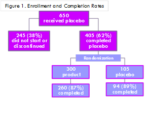
Childhood food allergy affects about 8% of US children. Recent research has revealed protective effects of early dietary introduction of allergenic foods on the development of food allergy for infants, including those at elevated risk. The goal of this study was to evaluate the safety and acceptability of a blend of 16 common allergenic proteins (peanut, soy, almond, cashew, hazelnut, pecan, pistachio, walnut, wheat, oat, milk, egg, cod, shrimp, salmon, and sesame) combined with 400 IU of Vitamin D into a food supplement powder. Caregivers were instructed to mix the powder into a solid or liquid feeding once a day. All procedures were deemed exempt by the Northwestern University IRB. A national sample of healthy infants, 5-11 months of age, without severe eczema participated in the 28-day placebo period followed by a 28-day randomized, blinded, placebo-controlled period. Caregivers were instructed to feed the infant one packet of the food supplement powder per day, observe their infant for 2 hours after ingestion, and record, in a web-based diary, any symptoms or allergic-type reaction including anaphylaxis occurring within 2 hours of ingestion and any reaction-related prescribed medication or medical care. Caregiver perceptions of the food supplement’s smell, texture, and packaging, were also assessed. Figure 1 shows enrollment and completion rates of the study. Of the 8,400 food supplement ingestions, no infants had any allergic reaction nor received any prescribed medication or medical care. Of 14,252 placebo ingestions, 1% (N=250) resulted and 0.7% (N=61) of food supplement ingestions in a report of symptoms (e.g., cough, diarrhea). This study suggests that the food supplement is safe and feasible for infants. Future study should assess the effect of the food supplement on immunologic responses to the allergenic proteins and on the incidence of food allergy

Abstract:

Jane L Holl is a General Pediatrician and Health Services and Outcomes Researcher who has conducted substantial prior research on childhood food allergy in the US. She is the Director of the Center for Healthcare Studies, an interdisciplinary center at Northwestern University. She has partnered previously with Kay Savio from Focus Pointe Global, Inc., a global market research company with fully vetted, precision-targeted participants

  • General Pediatrics | Neonatology | Pediatric Critical Care & Nursing | Pediatric Nutrition | Pediatric Trauma & Depression | Pediatric Endocrinology
Location: Malibu Room
Speaker

Chair

Jane L Holl

Northwestern University and Focus Pointe Global, Inc., USA

Speaker

Co-Chair

Kai Jiao

University of Alabama at Birmingham, USA

Session Introduction

Rania Abdelmonem Khattab

Cairo University, Egypt

Title: Common bacterial infections among children
Speaker
Biography:

Rania Abdelmonem Khattab has completed her PhD in 2012 from Faculty of Pharmacy, Microbiology and Immunology Department, Cairo University, Egypt. She has got Cairo University International Publication Award, Egypt in 2013, 2016 and 2017. She has many teaching experiences for both undergraduate and postgraduate courses e.g. basic microbiology and immunology, pharmaceutical microbiology, quality control of herbal drugs, public health and biotechnology. She has attended many workshops and some conferences with poster presentation in the Global Biotechnology Congress, Boston, MA, USA in June 2014 and 3rd Global Microbiologists Annual Meeting, Portland, Oregon, USA, 15-17 August 2016. She has given oral presentation in Clinical Trials Conference, Orlando, Florida, July, 2015. She has published many papers in international journals

Abstract:

Bacteria are tiny, microscopic, unicellular organisms. The most common bacterial infections among children are skin infections, ear infections, bronchiolitis, pinkeye (conjunctivitis), common cold and throat infections. Urine infection is common in children. It can cause various symptoms. A course of antibiotics will usually clear the infection rapidly. In most cases, a child with a urine infection will make a complete recovery. Certain children are at particular risk of bacterial infections. These children include: infants younger than 3 months, children who have no spleen or who have an immune system disorder and children who have cancer. Sometimes doctors diagnose bacterial infections by the typical symptoms they cause. Bacteria are identified in samples of tissue, blood, or body fluids, such as urine, pus, or cerebrospinal fluid. Bacteria from these samples can be identified under microscope, by culture or with a rapid identification test. Many bacterial infections in children can be prevented by vaccination. Viral infections such as measles, polio, and hepatitis A and B can also be prevented with routine immunization. Sometimes antibiotics alone can eliminate a bacterial infection. However, when an infection has created a large collection of pus, people sometimes also need surgery to drain the pus. Such infections include abscesses and joint infections. Infections spread among children by direct contact, indirect contact, droplets transmission and airborne spread. There are several ways to protect the child; by washing hands, using antibacterial hand sanitizers, cover his or her mouth when coughing or sneezing and clean open cuts

Carrie Owen

Childrens Hospital of Eastern Ontario, Canada

Title: Group success with the picky eater: Inter-professional parent group versus individual OT treatment

Time : 12:30-13:00

Speaker
Biography:

Carrie Owen is a Registered Occupational Therapist and Lactation Consultant specializing in infants, toddlers, young children, and teens with feeding challenges. She has provided expert advice and guidance to parents on subjects including development, feeding, and sensory processing. She is the Creator and Coordinator of the Inter-professional Feeding Groups for children between the ages of 5 months and 14 years at the Children’s Hospital of Eastern Ontario. She is presently studying the effectiveness of the feeding group for children between 4 and12 years of age. She was the Principle Author on a manuscript published in Nutrition in Clinical Practice and has also published in Today’s Parent magazine. Presently, she is working both at the Children’s Hospital of Eastern Ontario and in private practice. Her teaching experience includes the University of Ottawa, the University of Toronto and York University

Abstract:

Background: Feeding difficulties are common in young children and have been estimated to occur in 25-35% of typically developing children. The waitlist for occupational therapy feeding services has increased for numerous reasons including the new diagnostic category, avoidant/restrictive food intake disorder. To manage the waitlist, inter-professional feeding groups were offered. Although an inter-professional approach is recommended, the literature contains few systematic descriptions. In 2012, the Children’s Hospital of Eastern Ontario (CHEO) completed its program evaluation of this group approach and determined that it was an effective modality for children with feeding challenges. However, parents’ perceptions of this new approach are not well documented. The purpose of this study was to acquire early insight into parents’ perceptions of an inter-professional group (IG) service delivery model and an individual one-on-one (ONO) approach

Methodology: Participant eligibility was determined through referrals to occupational therapy for feeding challenges. 40 participants, (n=20 per group), were recruited using convenience sampling. Assignment was through simple randomization. The measuring process of care (MPOC)-20 was used to assess parents’ perceptions of healthcare professionals’ behavior

Findings: The IG scored high across all five constructs (mean range 5.1-5.9). Two significant constructs, coordinated and comprehensive Care and respectful and supportive care have means of 5.76 and 5.9 respectively. The scores across all constructs and between groups could be ranked identically. Children in both groups acquired adequate feeding skill levels. Wait times were lowered by 71%. The IG children required fewer follow-up visits than the ONO children. Results offer further evidence that an inter-professional-lead group program can provide care that brings the parents satisfaction. The authors agree that the value provided by the IG is in-line with patient and healthcare expectations

Break: Lunch Break 13:00-14:00 @ Atrium

Hamza Abbasi

Unterberg Childrens Hospital at Monmouth Medical Center, USA

Title: Symposium on Efficacy of standardized feeding protocol for preterm infants

Time : 14:00-15:00

Speaker
Biography:

Hamza Abbasiis a third-year Pediatric Resident at The Unterberg Children’s Hospital at Monmouth Medical Center. He has completed his MBBS from Alfaisal University School of Medicine in Riyadh, Saudi Arabia in 2014. He has been involved in multiple clinical research projects since 2011 and plans on continuing to play a role in optimizing health care via research in the future throughout his career as a Primary pediatrician

Abstract:

Although it is universally accepted that infants who are delivered prematurely require nutritional support to survive, the published evidence lacks clear indications of the best method of delivery, substrate use, timing or appropriate outcome measures to evaluate nutritional support. In the absence of published or widely accepted guidelines regarding nutritional support strategies for preterm infants, most institutions have developed their own guidelines for nutrition management and nutrition practices vary widely. We planned to assess the efficacy of a newly introduced standardized feeding protocol for preterm infants (<1800 g) in our hospital. These groups were stratified into 3 categories based on weight (i.e. <1000 g, 1000-1500 g and >1500 g). We calculated the number of days to regain birth weight and the number of days on TPN in both the groups to assess if there was any statistically significant difference between the pre-intervention and post intervention group. There was no statistically significant difference in the number of days to regain birth weight in any of the groups, and in the number of days on TPN noted in the <1000 g and >1500 g weight categories between the pre-intervention and the post- intervention group. However, there was a statistically significant difference (p<0.05) in the number of days on TPN in the 1000 g-1500 g weight category between the pre-intervention and the post- intervention groups. With this study, we can state that the implementation of a standardized feeding protocol in the NICU is efficient in terms of decreasing the number of days on TPN in preterm infants between 1000g and 1500 g

Seung Min Bae

Gachon University Gil Medical Center, South Korea

Title: Somatization and PTSD in child sexual abuse
Speaker
Biography:

Seung Min Bae completed a Psychiatric Training and Child Psychiatry Fellowship at Kangbuk Samsung Hospital. Since 2009, she has been a Psychiatric Assistant Professor at Gachon University and Director of Incheon Sunflower Center for Sexually Abused Children and is currently the Head of the Support Center for victims of crime in the ministry of justice. She is a non-standing Director of the Korea Women's Development Agency under the Ministry of Gender Equality and Family

Abstract:

Purpose: Somatization is a major post-traumatic symptom in the sexually abused children. The aims of this study were 1) to find the relation between somatizations and post-traumatic stress disorder (PTSD) symptoms in child sexually abused victims and 2) to elucidate whether type of abuse have effect on somatization.

Methods: Sixty-three victims were evaluated of their somatizations (Child behavioral checklist), PTSD symptoms (trauma symptom checklist for children, TSCC), and level of intelligence. The correlation and regression analyses were calculated to predict somatization based on the PTSD symptom, intelligence score, age, and the type of sexual abuse.

Results: The result showed that factors associated with somatization in the sexual abuse victims were PTSD symptoms (B=0.356, p=0.001) and intelligence (B=0.179, p=0.019). The type of abuse by itself did not correlate somatization (B=3.027, p=0.310), but had moderating effect of PTSD symptoms on somatization (Type of abuse PTSD symptoms, B=-.489, p=0.036). PTSD symptoms were associated with somatization only in victims with the molestation type of abuse.

Conclusions: Somatization of the sexually abused victims was influenced by the severity of PTSD symptoms and intelligence. The effect of the PTSD symptoms on somatization was mediated by the type of abuse. The rape type of abuse might be a powerful predisposing factor which attenuates the effect of post-traumatic symptoms on somatization

Speaker
Biography:

Hanaa I Rady is working as an Associate Professor of Pediatrics and Pediatric Intensive Care, and is the Deputy Director of Children University Pediatric Hospital, AbolRish, Cairo University

She is the Director of PICU of the 7th floor (21 beds). She has completed her MSc and MD from Cairo University in the year 2005 and 2008 respectively. She has published over 18 publications in reputed journals.

Abstract:

Background: Intensive care units (ICU) are always nearly full capacity which makes admission very difficult and may affect patient’s outcome at times of shortage.

Aim: To identify the variables capable of predicting which patient will benefit most from ICU admission, therefore prioritizing those patients when there is shortage of ICU beds.

Methodology: Two-hundred and fifty cases were subjected to thorough history taking, meticulous clinical examination, laboratory and radiological investigations when needed.

Results: ICU admissions occurred within 24 hours of coming to the emergency room (ER) in 63.6% of cases and after 24 hours in 22.4% of cases. Mortality rate was 37.6%. Survival was better among early than late (>24hrs) ICU admissions (p=0.05). Worst outcome was associated with pre-admission cardiac arrest (p<0.001), need for pre-admission intubation (p<0.001), high initial total inotropic dose (p<0.001) and thrombocytopenia (p<0.001).

Conclusion: Early ICU admission when indicated is associated with a better outcome. Pre-admission assessment can help prioritize those who would benefit from ICU. Not all cases referred to the ICU needed an intensive care, and most of them improved when received early care in the general ward.

Break: Coffee Break 16:00-16:20 @ Malibu Foyer
Speaker
Biography:

Showkat Hussain Tali is working as an Assistant Professor of Pediatrics, Adesh University. After obtaining his Bachelor's Degree in 2005, he obtained his MD in Pediatric Medicine from University of Kashmir in 2010. In 2013 he joined Department of Neonatology Surya Children’s Hospital Mumbai and became Board Certified in Neonatology from the National Board of India in 2016. In the same year he joined Adesh University as Assistant Professor Pediatrics and In-charge Neonatology. He has more than a dozen publications in national and international journals. He has received science talent search award from the Govt. of Jammu and Kashmir in 1997. He is also keenly interested in poetry and creative writing and has been awarded by Help Foundation and Rajiv Gandhi foundation, India, for excellence in creative writing in 2007

Abstract:

Aim: To compare the effect of 3-hourly (3-h) versus 2-hourly (2-h) feeding schedules on time to reach full feeds in neonates weighing less than 1500 grams

Materials & Methods: This was a randomized trial conducted in a level 3 neonatal intensive care unit, Surya Children’s Hospital Mumbai, India. We enrolled 120 preterm neonates with birth weights of 501 to 1500 g. The neonates were divided into 2 strata based on birth weight: 501 to 1000 g and 1001 to 1500 g. The neonates were randomized into 2 orogastric feeding schedules: 8 or 12 feeds (3-h or 2-h schedules, depending upon randomization), and a uniform feeding protocol was followed. Analysis was performed using the intention-to-treat principle. Categorical variables were compared using the Chi-square test. Continuous measures between groups were compared using 2-sample t test or Mann Whitney U test as appropriate. Data were analyzed using IBM SPSS version 21 software. P<0.05 was considered significant.

Primary Outcome measures were time (in days) to reach full feeds (defined as tolerance of 150 mL/kg/d of feeds for at least 48 h).

Secondary Outcome measures were time (in days) to attain birth weight; time (in days) to discharge; weight, length, and head circumference at discharge; incidence of feed intolerance, necrotizing enterocolitis (NEC), intravascular hemorrhage (IVH), screen-positive sepsis, culture-positive sepsis, hypoglycemia, apnea, jaundice and retinopathy of prematurity (ROP), duration of total parental nutrition (TPN) and nursing; and mortality.

Results: A total of 215 neonates were assessed for eligibility, of which 95 were excluded. Hence, 120 neonates were enrolled in the trial. There was no significant difference in time (in days) to reach full feeds in the 2-h versus 3-h groups (9.53±4.26 vs. 9.85±5.48; P=0.73). There was no significant difference between the 2 groups in any of the secondary outcomes. The total time spent per day in feeding was significantly lesser in the 3-h feeding schedule groups (P=0.04).

Subgroup Analysis revealed that among the neonates in the lower birth weight strata (501 to 1000 g), those fed 2 hourly reached full feeds earlier compared with those fed 3-hourly (2-h group: 11.24±2.88 d vs. 3-h group: 14.14±4.98 d; P=.041).

Conclusions: There was no significant difference in time to reach full feeds in all the neonates, irrespective of whether they were fed 2-h or 3-h. However, neonates < 1000 g reached full feeds earlier when fed more frequently (2-h feeding schedule)

Bahauddin Ibraheem Sallout

Womens Specialized Hospital- King Fahad Medical City, Saudi Arabia

Title: Can MCA Doppler studies predict mortality in fetuses with congenital hydrocephalus?
Speaker
Biography:

Bahauddin Ibraheem Sallout is a certified Maternal-Fetal Medicine (MFM) consultant from University of Ottawa, Canada, with American specialty in Ultrasound in Obstetrics and Gynecology. He has special training in fetal echocardiograph and 3D/4D sonography. He has developed the ultrasound unit and established the MFM department. Currently, he is the Medical Director for the Women’s Specialized Hospital, King Fahad Medical City. Riyadh, Saudi Arabia. He has 15 publications in the field of obstetrics ultrasound and fetal medicine, and he has participated and presented in many international and local conferences.

Abstract:

Objective: The objective of this study is to investigate the impact of abnormal middle cerebral artery (MCA) Doppler on the perinatal mortality in fetuses with congenital hydrocephalus (CH).

Methods: A prospective study of all fetuses with CH who delivered at our hospital over a period of 7 years. Data were obtained from the ultrasound, labor room and intensive neonatal care unit (NICU) database. The Perinatal mortality rates were evaluated in relation to the following measures, associated congenital anomalies, cortical mantle thickness (CMT), and MCA Doppler abnormalities (absent or reversed diastole). The main outcome measure was perinatal mortality rate in relation to MCA Doppler changes.

Results: A total of 85 cases of CH were diagnosed and managed. The birth prevalence of CH was 2.44 per 1000 live births. On one hand, the perinatal mortality rate was higher in those fetuses with non-isolated hydrocephalus, (37.25% (19/51) versus (35.29% (12/34, p¼0.854 and in those cases with CMT <10 mm, 38.78% (19/49) versus 33.33% (12/36) in those with CMT >10 mm, p¼0.607. On the other hand, the perinatal mortality rate was significantly higher in those fetuses with abnormal MCA Doppler, (100% (13/13) versus 25% (18/72), OR¼78.0, 95% CI (5.52–44085124.60), p<0.001.

Conclusions: Abnormal fetal MCA Doppler (absent or reversed diastole) appears to be a poor prognostic indicator with significantly high perinatal mortality in fetuses with CH

Day 2 :

Keynote Forum

Shoshana Dayanim

Keiser University, USA

Keynote: Infants, video viewing and learning

Time : 09:05-09:45

Biography:

Shoshana Dayanim earned an MA Degree in Creative Arts Therapy, and practiced as a Psychotherapist for several years before returning to school to earn her PhD in Applied Developmental Psychology from Fordham University and completing a Post-doctoral Fellowship at Emory University. Her research interests focus on the effects of television and technology on child development and ranges from infancy through adolescence. She has served as an Analyst and Research Consultant for various organizations involved in the development of educational media for children. She currently is the Chair of the IRB and Professor of Psychology at Keiser University

Abstract:

There is no question that children today are immersed in technology. They are exposed to video and technology from the youngest ages, even before they can hold a device. There is strong evidence that preschool and school aged children can learn from quality television programming, however, the research concerning infants 18 months and younger is limited, offers little evidence of possible benefits, and questions the adverse impact video may have on cognitive development. Despite the recommendations from the American Academy of Pediatrics that children younger than 18 months should refrain from screen media, with the exception of video chatting, videos targeted toward infants continue to saturate the market. Many of these videos purport to enhance infant vocabulary and language development. Because the use of baby signs enhances infants’ communicative repertoire and appears to serve the same communicative functions as words for young children, baby signs offer the opportunity to examine the efficacy of video instruction towards infant language development in a controlled experiment. This talk will provide a brief overview of the existing research on the impact of video viewing on infant cognitive development, and will focus on the findings of a four-week longitudinal experiment that investigated 15-month-old’s ability to learn american sign language signs from at-home viewing of instructional video, either with or without parent support, compared to traditional parent instruction and a no-exposure control condition. Forced-choice, elicited production, and parent report measures indicate learning occurred across exposure and testing conditions. This constitutes the first experimental evidence of infants’ ability to learn expressive communication from commercially available educational videos. These findings offer educators and physicians evidence based insights to help parents make the best choices available concerning infant screen time

Keynote Forum

Natasha Lepore

Childrens Hospital Los Angeles, USA

Keynote: Understanding the brain anatomy of premature and healthy newborns using MRI

Time : 09:45-10:25

Biography:

Natasha Leporé involves in the development of numerical tools for the analysis of brain magnetic resonance imaging data. She also works on applying these methods to different clinical and neuroscience applications. She is currently working as an Assistant Professor in Radiology at the University of Southern California and at Children's Hospital Los Angeles. She has Graduated with a BSc in Physics and Mathematics from the University of Montreal and then obtained a Masters in Applied Mathematics from Cambridge University. She has completed her PhD in theoretical physics (Harvard University), later she switched to neuroimaging and became a Postdoctoral fellow at the Laboratory of Neuro Imaging

Abstract:

I will cover some of the MRI analysis tools that we designed to understand the brain anatomy of newborns, such as cortical and subcortical morphometry and white matter tract analyses in these populations. I will also give some results on our analyses of early development of subcortical structures and networks in preterm and term born neonates and infants

Coffee Break 10:25 - 10:45 @ Malibu Foyer

  • General Pediatrics | Pediatric Oncology and Haematology | Neonatology | Pediatric Allergy & Infectious Diseases | Pediatric Endocrinology | Pediatric Cardiology | Pediatric Gastroenterology
Location: Malibu Room
Speaker

Chair

Shoshana Dayanim

Keiser University, USA

Speaker

Co-Chair

Showkat Hussain Tali

AIMSR, India

Speaker
Biography:

Nur Rochmah is working as a Lecturer of Department of Pediatrics, Faculty of Medicine, Airlangga University, Dr Soetomo Hospital. Surabaya. East Java. Indonesia

Abstract:

Background: Hypothyroidism is the common endocrine complications in thalassemia patients, caused by iron overload and iron depotition in thyroid gland. Early recognition and prevention may improve the quality of life. Thyroid dysfunction usually occured after 10 year in transfusion dependent thalassemia, unfortunately hypothyroidism is not clinically observed.

Objective: To determine the prevalence of hypothyroidsm pediatric thalassemia patients in Dr. Soetomo Hospital Surabaya

Methods: This cross sectional study evaluated children aged 3-18 years old with thalassemia at hematology-oncology outpatient clinic, Dr. Soetomo Hospital from February to March 2017. Inclusion criterias were: patients had regular transfusions more than one year. Clinical manifestations and biochemical analysis were taken. Subclinical hypothyroidism was defined as normal FT4 with high TSH; central hypothyroid: low FT4 and low to normal TSH; overt primary hypothyroidism: low FT4 and high TSH. Patients with hypothyroidsm were divided into 2 group, group 1(<11yo), and group 2(≥11yo).

Result: 106 patients were included in the study. 56 were male, mean age was 10.66 years. Moderate malnutrition was obsereved in 29.3%; mean of ferritin level was 3703.82±335.69; mean of age of start transfusion was 6.16. Mean of pre-transfusion hemoglobin level was 5.14±1.2. Impaired thyroid function were observed in 27 (25.2%) out of 106 patients: 12 (12.1%) patients subclinical hypothyroidism; 13 (13.1%)patients central hypothyroid, and 2 (2.0%)patients overt primary hypothyroidism. Comparison between group 1 and group 2 was significant(p<0.001).

Conclusion: Hypothyroidism is more prevalent in aged 11 year old and more. Thyroid function should be followed periodically

Speaker
Biography:

Kagiso Batka-Makwinja is a qualified medical doctor doing her fellowship specialization in Pediatric Surgery at the University of Pretoria. She serves in the executive committees of the South African Pediatric Surgical Trainees Association, the South African Society of Surgeons in Training and the International Pediatric Endoscopic Group. Dr Batka-Makwinja has presented topics globally: 'Infant Feeding in HIV Positive Mothers' in Barcelona, Spain; 'HIV Treatment Compliance' in Fortaleza, Brazil; Pediatric Laparoscopic Ovary-sparing Surgery in Benign Ovarian Neoplasms' in Kwazulu-Natal and 'Pediatric Laparoscopic Appendectomies' in Cape Town, South Africa. Kagiso Batka-Makwinja lives in Gauteng, South Africa

Abstract:

Gastroschisis has been used as the bellwether condition for assessing an institution’s capacity to deliver newborn surgical care. Therefore evaluating the treatment and outcomes of gastroschisis neonates is of importance to any unit offering neonatal surgical care. These neonates are particularly susceptible to sepsis, and mortality due to sepsis. The survival rate in developed countries has increased to 90%, but in developing countries may be as low as 20%.

This prospective study was aimed at determining whether implementation of and adherence to aseptic interventions could improve the survival rate of gastroschisis neonates, in this high prevalence region. The interventions included, written policies prioritising and re-enforcing hand washing and a ‘bare-below-elbows’ approach, establishing compliance to uniform blood culture taking techniques, using new Silo-bags for staged abdominal closures, and insertion and maintenance of central lines according to standardised protocols. Video material and physical demonstrations were used to demonstrate the techniques described in the above protocols. The World Health Organization’s (WHO) Hand Hygiene Self-Assessment Framework was used and this is a systematic tool to obtain situational analysis of hand hygiene promotion and practices in a specific unit. The survival rate of gastroschisis neonates increased to 67% from 40% after implementation and adherence to aseptic techniques and interventions. Compliance to aseptic protocols increased by 43%. Observations demonstrated a decrease in the rate of blood culture contamination rate. These findings may be utilised to drive initiatives and strategies to improve implementation policies at any level or setting of healthcare services

Costa Monteiro L M: Poster 1

National Institute of Women, Adolescent and Child Health Fernandes Figueira, Brazil

Title: Evaluation of urological outcomes in children exposed to fetal infection by Zika virus
Speaker
Biography:

Lucia M. Costa Monteiro, M.D, Ph.D., works at Instituto Fernandes Figueira/Fiocruz (www.iff.fiocruz.br), the Brazilian National Institute of Health for Women, Children and Adolescents. Her research interest is neurogenic bladder and voiding dysfunction and since 1998 has been the research group leader: (http://dgp.cnpq.br/dgp/espelhogrupo/3929782950316727).  From 2002-2004 she collaborated/worked as a research associated at the Children's National Medical Center. Member of the International Children's Continence Society and Member of Editorial Board Member of Editorial Board of Revista Brasileira de Saúde Materno Infantil (2006), Jornal de Pediatria (2008) and SM Journal of Nephrology and Kidney Diseases (2017).

Most recent publication: Early treatment improves urodynamic prognosis in neurogenic voiding dysfunction: 20 years of experience. J Pediatr (Rio J). 2017 Jul - Aug;93(4):420-427. doi: 10.1016/j.jped.2016.11.010. Epub 2017 Apr 19.

Abstract:

Background & Aim: Zika virus is a mosquito borne flavivirus that may affect pregnant women and their infants by causing fetal abnormalities. This Congenital Zika Syndrome (CZS) is associated with microcephaly and central nervous systems malformations. We identified that some of the regions damaged by CZS are also known to influence the neural circuitry controlling the lower urinary tract. The goal is to investigate an association of CZS and neurogenic urinary tract dysfunction to increase knowledge in the field and mitigate the impact of the disease in infected children.

Methods: Urological assessment was performed in pediatric patients with confirmed CZS referred to our Urology Clinic between June 2016 and June 2017. It consisted of clinical history, laboratory tests, renal ultrasound and urodynamic evaluation. ZIKV was previously confirmed by maternal history and positive PCR in babies/mothers. Microcephaly and other CNS abnormalities were established based on neurological and image evaluation (CT and/or MRI).

Findings: Twenty eight CZS patients were tested, 15 females and 13 males, age 10 months old in average (ranging from 2 mo to 19 mo old). All were presented with microcephaly. Urological assessment confirmed neurogenic urinary tract dysfunction in 100%, some with high risk profile. Most were asymptomatic but urological screening confirmed urinary tract infection in 5 and renal US were already abnormal in 3 patients.

Conclusion: Urological comorbidities are associated with congenital Zika syndrome, including high risk urodynamic patterns that can cause renal damage if left untreated. This is potentially the only treatable health condition in CZS setting and neonatologist and pediatricians need to be aware to promote proactive management that mitigates disease burden to patients and their families.

Speaker
Biography:

Shagufta Yousuf is working as Assistant Professor at Adesh University, India. After obtaining her Bachelor's degree in 2007, she obtained her Post-graduate Diploma in Maternal Child Health from IGNOU in 2011 and MD in OBG from University of Kashmir in 2016. In the same year she joined Adesh University as Assistant Professor, OBG. She has several publications in national and international journals. Recently she has been invited to deliver a speech at International Congress of Gynecology and Obstetrics, Taiyuan, China (Nov 2017)

Abstract:

This near term, 36+4 weeks, 2.8 Kg birth weight, AGA, non con-sanguinous product, male infant was admitted to Surya Child Care Neonatal Intensive Care Unit (SCH-NICU) on day 23 of life with respiratory distress since birth. Infant was born to a 32 years G2P1L1 mother. Antenatal period was uneventful. It was an LSCS delivery for non-reassuring fetal status. Baby was vigorous at birth and resuscitation was not required. However, infant was noticed to have respiratory distress soon after, for which infant was admitted to an NICU at Indore, India. At admission infant was given one dose of surfactant for suspected respiratory distress syndrome. Infant was ventilated up to day 8 of life, given one more dose of surfactant at day 8 of life, extubated and put on oxygen supplementation through nasal prongs. Infant was reintubated at day 11 of life for increasing respiratory distress. Chest X-rays showed persistent bilateral haziness. Infant was given multiple antibiotics with suspicion of congenital pneumonia. However there were no significant antenatal risk factors for sepsis and infant’s sepsis work-up was unremarkable. CT chest showed patchy areas of air space consolidation bilaterally. As respiratory distress was persistent, infant was transferred on 23 day of life to Surya Child Care NICU, Mumbai for further care. On admission, infant was put under ventilation support for significant respiratory distress. Differential diagnoses was considered which included; unresolved pneumonia; congenital heart disease (TAPVC); GERD; H type tracheo-esophageal fistula; immunodeficiency; cystic fibrosis; alpha 1 antitrypsin deficiency; congenital pulmonary alveolar proteinosis; congenital lymphangeictasia; primary ciliary dyskinesia and CMV pneumonitis. Respiratory distress persisted throughout the admission. Chest X-rays performed periodically showed persistent haziness. Ventilation assistance was required throughout the admission. Serial sepsis screens and blood cultures didn’t show any evidence of sepsis. CSF study, tracheal secretion cultures, CMV antibody titers and CMV DNA-PCR were all non-remarkable. Work-up for immunodeficiency including flow cytometric lymphocyte sub-set analysis was unremarkable. 2D echoe of the heart, cranial ultrasound and USG of abdomen and pelvis were normal. Milk scan revealed presence of high grade (IV) gastro esophageal reflex. Repeat milk scan after fundoplication showed no evidence of GER. Infant’s HRCT revealed diffuse ground glass opacities bilaterally suggestive of alveolar edema of uncertain cause with normal tracheo-bronchial tree. Work-up for cystic fibrosis (delta 508 mutation) and alpha 1 antitrypsin deficiency were unremarkable. Electron microscopy of nasal scraping for cilia morphology was unremarkable. Bronchoalveolar lavage revealed lipid laden macrophages but little PAS positive staining. Histopathological examination of the excisional lung biopsy revealed PAS positive material in alveolar spaces with preserved alveolar architecture. Electron microscopy examination was suggestive of congenital alveolar proteinosis. For financial constraints further evaluation could not be performed. Despite giving optimal supportive care, performing partial broncho-alveolar lavage (once), administering IVIG, methylpredinosolone, and granulocyte colony stimulating factor, no significant response was observed. On request, infant was transferred back to Indore and died peacefully in an NICU after 3 days of transfer.

Speaker
Biography:

Background: Obesity in children are increasing rapidly lately. This may lead to insulin resistance and Diabetes Mellitus in adulthood. Acanthosis nigricans is manifestation of insulin resistance in the skin. The relationship between Acanthosis Nigricans(AN) and impaired glucose tolerance in overweight and obese children are still controversial.

Objective: To analyze relationship between acanthosis nigricans and impaired glucose tolerance in overweight and obese children.

Methods: This cross-sectional study was conducted between February until March 2017 in Pediatric endocrine outpatient clinic and Pediatric ward Dr. Soetomo Hospital. The presence of acanthosis nigricans was verified by Pediatric Endocrinology consultant, anthropometric and waist circumference measurements were taken. Oral glucose tolerance test was performed. Inclusion criterias were patient age 3-18 years old and BMI. Exclusion criterias were patient wih severe condition or in PICU and with congenital syndrome. Statistical analysis was using comparative study. Data analysis were performing using the SPSS 17.

Results: A total of 30 children were studied, 53.3% were boys, mean of age 10 year, mean of BMI 23.4, mean of waist circumference 87.6+/-12.8 cm. There were 21 patients had acanthosis nigricans and 14 with impaired glucose tolerance. Five obese children showed severe, seven with moderate and five with mild neck acanthosis nigricans respectively. Obese children were more likely to have acanthosis nigricans (P=0.019). There was significant correlation between acanthosis and impaired glucose tolerance (P=0.032).

Conclusion: There was relationship between acanthosis nigricans and impaired glucose tolerance in obese and overweight children.

Abstract:

Nur Rochmah is working as a Lecturer of Department of Pediatrics, Faculty of Medicine, Airlangga University, Dr Soetomo Hospital. Surabaya. East Java. Indonesia

Speaker
Biography:

Showkat Hussain Tali is working as Assistant Professor Pediatrics, Adesh University. After obtaining his Bachelor's degree in 2005, he obtained his MD in Pediatric Medicine from University of Kashmir in 2010. In 2013 he joined Department of Neonatology at Surya Children’s Hospital, Mumbai and became Board Certified in Neonatology from the National Board of India in 2016. In the same year, he joined Adesh University as Assistant Professor Pediatrics and In-charge Neonatology. He has more than a dozen publications in national and international journals. He has received Science Talent Search Award from the Govt. of Jammu and Kashmir in 1997 and has been awarded by Help Foundation and Rajiv Gandhi Foundation, India, for excellence in creative writing in 2007. On May 26/2017, he presented a speech at International Congress of Gynecology and Obstetrics, Prague, Czech Republic and has been invited to deliver speech at International Congress of Pediatrics, Taiyuan China (Nov 2017)

Abstract:

A full term, male infant with no significant antenatal and birth history developed severe respiratory distress on day 2 of life. Infant was diagnosed to have H-type of tracheoesopheageal fistula (TEF) and was operated for the same on day 4 of life. Infant was extubated on day 20 of life (difficult extubation) and was put on HHHFNC (heated humidified high flow nasal cannula). Soon after extubation, infant developed severe respiratory distress and stridor. Infant was put back under ventilator support. Flexible laryngoscopy along with bronchoscopy was performed under light sedation. Except for mild subglotic edema, no abnormality was detected. Size 3.5 ET (endotracheal) tube was replaced with a 3 size ET tube and a short course of dexamethasone (0.2 mg/kg/day × 5 days) was administered. After a 10 days period, the infant could be weaned to CPAP (continuous positive airway pressure). However it was not possible to take the infant off the CPAP thereafter. CECT (contrast enhanced computed tomography) was performed and no significant abnormality was detected. Parents were counseled for a tracheostomy but they refused. After one month period, when there was no improvement in clinical condition, laryngoscopy with bronchoscopy was again performed under anesthesia. Tight aryepiglotic folds were detected and aryepiglotic split was performed. Infant responded dramatically to treatment and could be weaned to room air within 3 days of surgery. The anesthesia technique has been found to be superior to awake technique with a sensitivity, specificity, positive predictive value and negative predictive value of 100% each as compared with 93%, 92%, 97%, and 79%, respectively, for awake technique. Most probably, we missed the diagnoses in the first place as we didn’t perform the laryngoscopy under anesthesia or sufficient sedation. It is worth mentioning that laryngoscopy along with bronchoscopy and esophagoscopy was performed under anesthesia during the initial evaluation of TEF before surgery. This makes us strongly believe that the tight aryepiglotic folds were a complication of TEF repair surgery or prolonged intubation rather than a congenital one

Speaker
Biography:

Mohamed Almuqamam has completed his MD at the age of 23 years from the Royal College of Surgeon in Ireland (RCSI-Bahrain). He is a graduate of the UK Foundation School in Malta 2014-2016 and is currently a PGY2 Pediatric resident at The Brooklyn Hospital Center

Abstract:

Introduction/Objectives: Acute pain episode is a common reason for patients with sickle cell disease to present to the ER. This study is designed to assess the role of multiple factors that might affect the time from ED triage to the administration of the first opiate pain medication and its dosage, to compare current practices with the American Pain Society Guideline for the Management of Acute and Chronic Pain in Sickle-Cell Disease in the emergency department. By identifying and recognizing some of the factors that delay or affect the proper dosing of the pain medications, we aim to implement suitable and plausible changes to ensure better emergency care for these sickle cell disease patients.

Methodology: This is a cross-sectional descriptive study that relied on collecting non-identifiable data from the local EMR to assess for possible relationship between the proposed set of factors/variables and the time to administration of the 1st narcotic pain medication and it’s dosage. The population in question includes the entire sickle cell disease patients’ population (HB-SS, HB-SC, HB-SD, HB-SB+ and HB-SB0) that are under the care of our Pediatric Heme-Onc clinic at The Brooklyn Hospital Center (TBHC) with the age range of 1 day to 21 years. The factors include age, gender, pain assessment/scale, time of presentation, mode of arrival, presence or absence of IV access at presentation, and ESI acuity. SPSS program was used for statistical analysis and treatment with a pre-set P-value at 0.05.

Results: There were 259 patient ER visits with 148 unique patients. Mean (SD) age of the entire study population was 15.98 (+/- 4.08) years and 61.8% of the patients were females. Average time to 1st opiate pain medication was 120.27 minutes (SD +/- 78.4) and average doses of Morphine and Hydromorphone were 0.067 mg/kg and 0.053 mg/kg respectively. Longer waiting time to 1st opiate pain medication were found in females with a mean difference of 25.5 minutes (95% CI 20 – 80.5 P value 0.027), older patients and patients with least severity ESI score (correlation coefficient of 0.214 & 0.134 (p values of 0.001 and 0.031) respectively). On the other hand, there seems to be a negative correlation between the time to 1st opiate and the pain score with a negative correlation coefficient of -0.22 ( p value of <0.001).

Conclusion: Overall, patients with acute SCD pain experienced significant delays when seeking pain relief in the ED. The following patients experienced the longest delays: those assigned a lower triage priority level, female patients, patients with lower pain score and older patients

Speaker
Biography:

Ahsan Akhtar is a PGY-2 Pediatric Resident at The Brooklyn Hospital Center. His interest is primarily in Neonatology especially placental disorders and new born outcomes. His focus was mainly to measure the effect of preeclampsia in newborns in the presence of low birth weight and very low birth weight. His other interests are respiratory modalities, nutrition including total parental nutrition and sepsis in neonates

Abstract:

Hypertension (HTN) disorders are the most common medical complication occurring in 12-22% of all pregnancies. Preeclampsia is responsible for about 15.9% of maternal deaths in USA. We hypothesized that early onset pre-eclampsia has greater impact on morbidity and mortality. Electronic medical records from January 2014 to December 2015 of infants birth weight (BW)<1500g were reviewed. Patients with co-morbidities were excluded. We identified 20 cases with HTN and preeclampsia, 69 healthy controls. The demographics, mode of ventilation, length of stay (LOS), morbidity and mortality were analyzed with Chi-square test. The Mann Whitney test was used to analyze the duration on different modes of ventilation. A p value <0.05 was considered statistically significant. No difference was noted in the maternal demographic data or laboratory values. No significant difference was noted in mode and duration of respiratory support, mortality rates, LOS, vasoactive support, blood products, retinopathy and brain abnormalities. The results were stratified to compare BW<1000 g and 1000-1499 g and continued to show no statistical difference in any of the measured outcomes. Comparing cases with BW<1000 g vs. 1000-1499 g, the mode and duration of respiratory support was significantly longer in <1000 g. LOS, vasoactive support, blood products and brain abnormalities were all higher in the <1000 g group. Comparing the controls with BW<1000 g vs. 1000-1499 g, LOS and mortality rate were higher in the <1000 g group. No differences were noted in vasoactive support, blood products and brain abnormalities. We found no significant difference in adverse outcomes between the groups. Even after further sub-categorizing the subjects based on BW, there still was no statistical difference between the groups. Infants born at a lower BW are believed to have more adverse outcomes after birth. Very low BW has a greater chance of morbidity and mortality compared to low BW infants if born to mother with hypertension

Break: Lunch Break 12:45-13:45 @ Atrium
Speaker
Biography:

Dr. Sallout is a certified Maternal-Fetal Medicine (MFM) consultant from university of Ottawa, Canada, with American Specialty in Ultrasound in Obstetrics and Gynecology. He has special training in fetal echocardiograph and 3D/4D sonography. He developed the ultrasound Unit and established the MFM department, and currently, the Medical Director for the Women’s Specialized Hospital, King Fahad Medical City. Riyadh, Saudi Arabia. He has 17 publications in the field of obstetrics ultrasound and fetal medicine, and he participated and presented in many international and local conferences

Abstract:

The prevalence of major congenital anomalies in Saudi Arabia is a largely understudied area. Knowing the prevalence of birth defects and their trends is important in identifying potential factors that are either causative or preventative. Early antenatal diagnosis of major congenital anomalies is important for possible termination of pregnancy, fetal or neonatal. We determined the prevalence of major congenital anomalies in our hospital population since implementation of an improved screening system.

This single-centre prospective cross-sectional study was conducted in a tertiary care hospital in Riyadh. A total of 63,452 obstetrical ultrasound examinations were performed for 30,632 female Saudi obstetric patients from the period of January 2007 to December 2012.

A total of 1,598 fetuses were diagnosed with major congenital anomalies, including 1,064 (66.6%) fetuses with isolated major anomalies and 534 (33.4%) fetuses with non-isolated major anomalies. The antenatal prevalence of congenital anomalies was 52.1 per 1000 pregnancies. The median maternal age at diagnosis was 29 years. The median gestational age at diagnosis was 30 weeks of gestation. Two hundred and eighty five cases (17.85%) had a previous family history of similar anomalies. The most commonly diagnosed anomalies involved the genitourinary system (652 cases). The birth prevalence of major congenital anomalies was 46.5 per 1000 live births.

The prevalence of major congenital anomalies in our hospital population appears to be higher than international prevalences, with a high recurrence rate. Environmental, nutritional and social factors may be contributing to this phenomenon

Speaker
Biography:

Muhammad Faizi is currently working as Lecturer at the Department of Pediatrics, Faculty of Medicine, Airlangga University- Dr Soetomo Hospital. Surabaya, Indonesia

Abstract:

Background: Mostly, type 1 diabetes mellitus (T1DM) is caused by pancreatic beta cell damage by autoimmune process which leads to reduced insulin production. Autoantibodies related to T1DM are islet-cell antibodies (ICA), insulin autoantibodies (IAA), tyrosine phosphatase autoantibodies (IA-2A) and glutamic acid decarboxylase (GAD). The profile of anti-glutamic acid decarboxylase autoantibodies in type 1 diabetes mellitus children in Dr. Soetomo Hospital Surabaya is still unknown.

Objective: The objective of the study is to determine the profile of anti-glutamic acid decarboxylase autoantibodies in type 1 diabetes mellitus children in Dr. Soetomo Hospital Surabaya.

Methods: This cross sectional study evaluated children with T1DM in Pediatric Endocrinology Outpatient Clinic of Dr.Soetomo Hospital during January-March 2017. We diagnosed T1DM based on consensus of Pediatric Endocrinology Working Group, Indonesian Pediatric Society. The presence of anti-GAD in serum was detected by anti-GAD65 commercial kit. Duration of illness referred to time from T1DM diagnosed until anti-GAD examination.

Results: There were 32 children with T1DM, 50% males. The mean age at diagnosis was 9.75 (SD 2.52, range 4-14) years. The median of duration of illness was 3.5 (range 1-11) years. The anti-GAD was positive in 30 (93.8%).

Conclusions: Anti-GAD test is positive in majority children with T1DM and still persist for long periods since T1DM diagnosed

Speaker
Biography:

Li-Xiao Xu is working as an Assistant Professor of Clinical Research Institute of Soochow University. She got her Doctorate degree in Biochemistry and Molecular Biology at Sun Yat-Sen University in 2012. Since 2012, she has been working in Children’s Hospital of Soochow University and focused on the research of brain damage, studies on the mechanisms of autophagy in hypoxia-ischemia-induced brain damage. Currently, she has hosted a National Natural Science Foundation-Youth Foundation (No.81502157), and obtained awards from Jiangsu Provincial Medical Youth Talent (No. QNRC2016758) and applied for Foundational Research of Medical and Health Care of Suzhou City (No.SYS201646). She also have been published many papers

Abstract:

Autophagy has been implicated to mediate experimental cerebral ischemia/reperfusion-induced neuronal death; the underlying molecular mechanisms, though, are poorly understood. In this study, we investigated the role of autophagy in regulating the expression of AMPAR subunits (GluR1, GluR2, and GluR3) in oxygen glucose deprivation/reperfusion (OGD/R)-mediated injury of hippocampal neurons. Our results showed that, OGD/R-induced hippocampal neuron injury was accompanied by accumulation of autophagosomes and autolysosomes in cytoplasm alongside a dramatic increase in expression of autophagy-related genes, LC3 and Beclin 1 and increased intracellular Ca 2+ levels. Pre-treatment with autophagy inhibitor 3-methyladenine (3-MA) significantly reduced this effect. Moreover, the OGD/R-induced up-regulation of mRNA and protein expressions of GluR1, GluR2 and GluR3 were also effectively reversed in cells pretreated with 3-MA. Our findings indicate that OGD/R induced the expression of GluRs by activating autophagy in in vitro cultured hippocampal neurons, which could be effectively reversed by the administration of 3-MA

Break: Coffee Break 15:45-16:05 @ Malibu Foyer
Biography:

Jing Liu is the Leader and the Director of the Center of Neonatal Intensive Care Unit, Beijing Chaoyang District Maternal and Child Health Care Hospital and Bayi Children’s Hospital Affiliated With the Army General Hospital of the Chinese PLA.His research interest is neonatal critical care, his academic positions includes the Associate Chairman of Committee of PLA Academy of Pediatric, the Associate Chairman of Neonatal Neurologist Committee of Chinese Neonatologist Association and Editorial Member of 20 Chinese or English Journals,etc. He has published over 260 papers as a First Author, 10 Books and Chapters in Books. His research work has been supported by China Natural Science Foundation and China Post-doctoral Science Foundation, etc., and he has won 12 awards for Science and Technology of The Government of China

Abstract:

Background: Ultrasound has been used extensively for the diagnosis of lung diseases including in neonatal intensive care unit (NICU). We have developed this technique in NICU for the diagnosis and differential diagnosis of neonatal lung diseases since January, 2011. Till now, a total of 6184 infants were accepted more 20,000 times lung ultrasound examinations. We found that almost all kinds of lung diseases can be diagnosed accurately by lung ultrasound.

Methods: Generally, the diagnoses of lung diseases were based on medical history, clinical manifestation, laboratory examination and the findings of chest radiography (CR) and/or computed tomography (CT). The ultrasonographic features of different lung diseases were compared with above traditional findings.

Results: There were 1597 cases without lung disease and 4587 cases were diagnosed as different lung diseases, including transient tachypnea of newborn (TTN) 1296 cases, respiratory distress syndrome (RDS) 1104 cases, pneumonia 1056 cases, meconium aspiration syndrome (MAS) 429 cases, pulmonary atelectasis 242 cases, pulmonary hemorrhage 107 cases, pneumothorax 86 cases, pulmonary edema 242 cases (who couldn’t be included to other diseases), 25 infants  were misdiagnosed as atelectasis  in fact  that they were thymus gland. Different lung disease has different ultrasonographic characteristics, generally, however, the main signs of lung disease on LUS were as following: pleural line abnormalities, absence of A-lines, lung consolidation with air bronchograms, interstitial syndrome, lung sliding disappearance, lung pulse, lung point,double-lung point, compact B-line dan white lung.

Conclusion: Ultrasound is advantageous in diagnosing neonatal lung diseases because of the following benefits: low cost, easy to learn and operate, non-invasive, harmless to the human body, and suitable for ongoing monitoring. Ultrasound is accurate and reliable and thus has an important value in guiding treatment and can replace X-ray as the preferred imaging method of neonatal lung diseases

Biography:

Dr. Hylemariam Mihiretie is currently working in Wollega University, Ethiopia. His research interests are pediatric HIV/AIDS, maternal and neonatal healthcare etc

Abstract:

Background: Intestinal parasites (IPs) are major concerns in most developing countries where HIV/AIDS cases are concentrated and almost 80% of AIDS patients die of AIDS-related infections. In the absence of highly active antiretroviral therapy (HAART), HIV/AIDS patients in developing countries unfortunately continue to suffer from the consequences of opportunistic and other intestinal parasites. The aim of the study was to determine the prevalence of intestinal parasites in relation to CD4+ T cells levels and anemia among HAART initiated and HAART naïve pediatric HIV patients in a model ART center in Addis Ababa, Ethiopia.

Methods: A prospective comparative cross-sectional study was conducted among HAART initiated and HAART naive pediatric HIV/AIDS patients attending a model ART center at Zewditu Memorial Hospital between August 05, 2013 and November 25, 2013. A total of 180 (79 HAART initiated and 101 HAART naïve) children were included by using consecutive sampling. Stool specimen was collected and processed using direct wet mount, formol-ether concentration and modified Ziehl-Neelsen staining techniques. A structured questionnaire was used to collect data on socio-demographic and associated risk factors. CD4+ T cells and complete blood counts were performed using BD FACSCalibur™ and CELL-DYN 1800, respectively. The data was analyzed by SPSS version 16 software. Logistic regressions were applied to assess any association between explanatory factors and outcome variables. P values <0.05 were taken as statistically significant.

Results: The overall prevalence of IPs was 37.8% where 27.8% of HAART initiated and 45.5% of HAART naive pediatric HIV/AIDS patients were infected (p<0.05). Cryptosporidium species, E. histolytica/dispar, Hook worm and Taenia species were IPs associated with CD4+ T cell counts <350 cells/μL in HAART naive patients. The overall prevalence of anemia was 10% in HAART and 31.7% in non-HAART groups. Hook wormS. stercoralis and H. nana were helminths significantly associated with anemia in non-HAART patients [AOR, 95% CI: 4.5(1.3, 15.2), P< 0.05]. The prevalence of IPs in non-HAART patients was significantly associated with eating unwashed/raw fruit [AOR, 95% CI: 6.3(1.2, 25.6), P<0.05], open field defecation [AOR, 95% CI: 9.3(1.6, 53.6), P<0.05] and diarrhea [AOR, 95% CI: 5.2(1.3, 21.3), P<0.05]. IPs significantly increased in rural residents [AOR, 95% CI: 0.4(0.1, 0.9, P<0.05)].

Conclusion: The overall prevalence of intestinal parasites significantly differed by HAART status and cryptosporidium species were found only in HAART naïve patients with low CD4+ T cell counts. Anemia was also more prevalent and significantly associated with IPs in non-HAART patients. This study identified some environmental and associated risk factors for intestinal parasitic infections. Therefore, Public health measures should continue to emphasize the importance of environmental and personal hygiene to protect HIV/AIDS patients from infections with intestinal parasites and maximize the benefits of HAART.